- Centro Interuniversitario per la Promozione dei Principi delle 3R nella Didattica e nella Ricerca
Microfluidics for 3D Cell and Tissue Cultures: Microfabricative and Ethical Aspects Updates

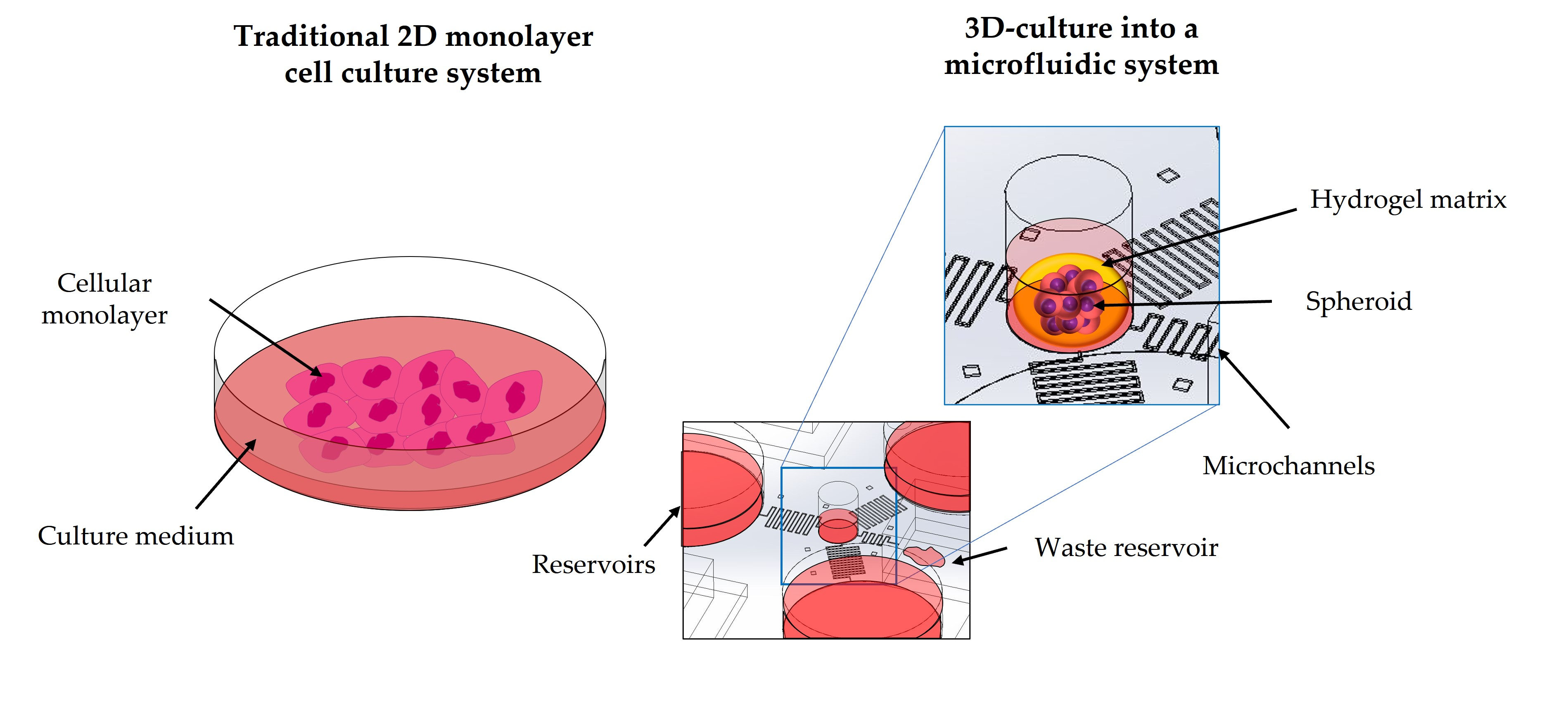
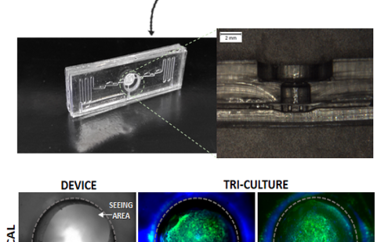
The necessity to improve in vitro cell screening assays is becoming ever more important. Pharmaceutical companies, research laboratories and hospitals require technologies that help to speed up conventional screening and therapeutic procedures to produce more data in a short time in a realistic and reliable manner. The design of new solutions for test biomaterials and active molecules is one of the urgent problems of preclinical screening and the limited correlation between in vitro and in vivo data remains one of the major issues. The establishment of the most suitable in vitro model provides reduction in times, costs and, last but not least, in the number of animal experiments as recommended by the 3Rs (replace, reduce, refine) ethical guiding principles for testing involving animals. Although two-dimensional (2D) traditional cell screening assays are generally cheap and practical to manage, they have strong limitations, as cells, within the transition from the three-dimensional (3D) in vivo to the 2D in vitro growth conditions, do not properly mimic the real morphologies and physiology of their native tissues. In the study of human pathologies, especially, animal experiments provide data closer to what happens in the target organ or apparatus, but they imply slow and costly procedures and they generally do not fully accomplish the 3Rs recommendations, i.e., the amount of laboratory animals and the stress that they undergo must be minimized. Microfluidic devices seem to offer different advantages in relation to the mentioned issues. This review aims to describe the critical issues connected with the conventional cells culture and screening procedures, showing what happens in the in vivo physiological micro and nano environment also from a physical point of view. During the discussion, some microfluidic tools and their components are described to explain how these devices can circumvent the actual limitations described in the introduction.
| Allegato | Dimensione |
|---|---|
| 1.57 MB |


